Pages

Monday 11 November 2019

Living Organisms - 1&2

WALT: Place organisms on a continuum
Part.1

Part.2

Task Description:
For this task room 4 and I were to complete the task of knowing what was smallest - largest. Part 1 explains the living organisms from smallest to largest. Part 2 explains the connections between the cells from smallest to largest. Overall this task was incredibly interesting as it made me actually want to lean about living organisms and our cells.   

Wednesday 6 November 2019

Dna - note taking!

Dna - note taking!

Today Room 4 watched a short video about cells and how they’re connected. Below this
introduction is my what I’ve been taking notes of. Learning more about cells and how they’re
connected was incredibly interesting. I really hope you enjoy! 
  
How is Dna, chromosomes and gene connected? Today we will be talking about how Dna,
chromosomes and genes relate. 


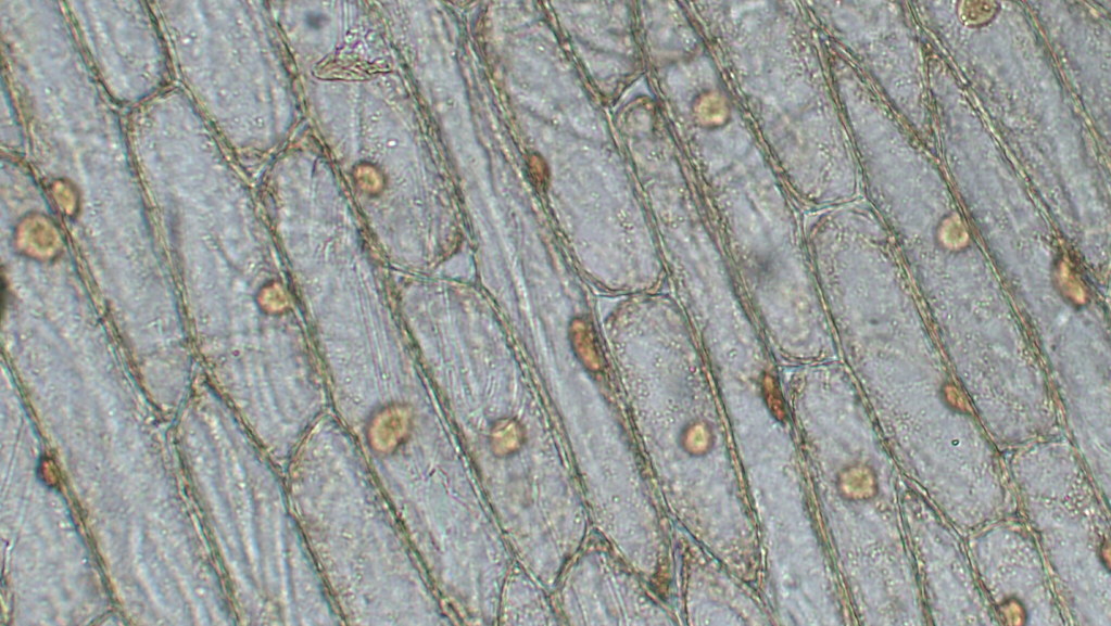
Our body is made up of hundreds and thousands of cells. Cells are only seen under a microscope. 


Humans have 23 pairs of chromosomes. Chromosomes have different sorts of information about
dna.


 Dna is made up of 4 basis (AKA) 4 different things. A,G,C,T. Dna is in the chromosome, the
chromosome is in the nucleus, and the nucleus is in the cell. 


The reason why Dna is shaped as a ladder is because…
A always joins with T  and G always joins with C


When a sequence of three pairs come together they create a word, then when we add more
words together we then create a chapter which is called gene.  


Gene’s are like chapters in a book. More than one or two genes is what creates our chromosome.
Chromosome is what lives in the nucleus.


Protein base letters:
Codons = words
Gene = Chapter
Chromosome = Book

Monday 4 November 2019

Science Experiment

Science Experiment!

Aim: see onion cells on a microscope


Hypothesis:
I think the 100x magnification we’ll be able to see what’s about to happen!

Equipment
Microscope Onion
Slide
Cover slip
Tweezers 
Blue dye - Blue stain


Method:
Our very first step started off with using a slide, after that we got ourselves a pair of tweezers and one onion, using the tweezers and onion we were to peel a thin layer of onion and gently placed it onto the slide, after placing the thin layer of onion onto the slide we were to pour a few drops of saline on top and add a bit of blue stain. The last step was to add a cover slip as it’ll help to make our vision more clear to see.


Observation
On the 10x microscope we were able to see the basic shape of the cell.
On the 40x microscope we were able to see a closer version of the segmented area.
On the 100x microscope we were only able to see the blue stain but from what our vision was the colour was purple.    

https://live.staticflickr.com/4164/34062338020_82b3009b48_b.jpg

Image attribution: